


Searching for our sense of self in the brain: are we on the right track?
#9

Investigating the brain mechanisms of our sense of self is misguided according to György Buzsáki.
Buzsáki is a highly respected neurophysiologist; professor at NYU; an elected member of the U.S. National Academy of Sciences; recipient of many honors and prizes. He has been active in systems neuroscience research since the 1970s and continues to produce cutting-edge work. Much of his research, along with that of many others around the world1, has focused on understanding how the hippocampus (a brain structure) is involved in complex tasks like forming long-term memories or navigating through our natural habitat.
Here is a link to Buzsáki’s website. More on his work later.
Extrapolating from Buzsáki’s published views2, the átman, rooh, psyche, ego - the ‘sense of self’ - is a concept we inherit from religion / philosophy going all the way back to ancient civilizations (Hindu, Islamic, Greek and such). It is part of a set of concepts like attention, imagination, instinct, free will, all of which are in everyday use, more or less. They were adopted by psychologists to describe our mental capabilities underlying our subjective experiences3.
Brains, on the other hand, starting with the neural nets of jellyfish, originated over 500 million years ago and have evolved since then. They are very much a product of evolution and subject to natural selection.
A long-standing and influential opinion amongst many neurophysiologists holds that brains evolved to help animals move about in their environment. Immobile, multicellular organisms (trees, for example) don’t need brains, and consequently don’t have them. Brains evolved, and continue to evolve, to allow mobile, multicellular life forms survive and reproduce while facing unexpected dangers and inconsistent opportunities that arise as they move about in their individual habitats4.
Nothing in nature directly corresponds to familiar philosophical and psychological concepts (e.g. language or musicality or even mathematical ability). These concepts with their culturally defined boundaries are unlikely to align with the real but unknown brain functions that underlie them and have evolved over geologic time. Nothing then, prevents these psychological constructs from either sharing brain mechanisms or having partially overlapping ones with no clear cut boundaries. Nature is blind to these artificial divisions of brain function imposed by humans. This mismatch between psychology and neurology may be referred to as the mind-brain mechanisms mismatch problem.
To clarify this mismatch, let us use a favorite analogy of some thinkers in this field. Consider the apps on your smart phone.

Apps have user friendly interfaces and deal in high-level concepts. For example, your fitness app calculates calories burned during a certain time period by measuring heart rate, counting steps etc. The various apps and their colorful charts and graphs are like emotions, memory, imagination etc. in this analogy.
Now consider the software code: it is probably written in Python or Java or some such high-level language underneath the user-friendly interface. Dig even deeper and you come to the hardware: the processor chips, integrated circuits, sensors etc. Which chip corresponds to the music app? Which one for the fitness app? We understand that the phone doesn’t really have specific chips for individual apps. The phone’s hardware is analogous to neural circuits and the molecular machinery of brain cells. The smart phone can make the charts for a fitness app, or alternatively graphs for a stock market app using the exact same machinery; much of the code is common as well. Our brain may be doing the same thing for attention, love, hate, greed, sense of self and many other psychological concepts that seem so distinct to us.
Our analogy breaks down very quickly though. Unlike a computer or a smartphone that needs to be filled with its operating system and apps only after it is fully built, the brain is a self-organizing structure. It is electrically active at the earliest stages of its embryonic development. Indeed, this spontaneous electrical activity is considered crucial for brain development, and is prominent before the brain begins receiving any signals from the sensory organs. Disrupting this spontaneous activity during embryonic development leads to abnormal development and consequent neurological and psychiatric disorders5.
Below is an excerpt from a recent review on brain development (referenced in footnote 5). The review summarizes recent discoveries on how neuronal networks of the brain’s cortex develop. (The cortex is the outermost layer of the brain thought to be involved in its most complex functions and has about 17 billion neurons (~20% of total) in humans; the thalamus is another brain region involved in relaying sensory inputs to the cortex)
… Patterns of spontaneous activity during early development synchronize local and large-scale cortical networks (made up of billions of neurons with trillions of synapses (connections) between them), which form the functional template for generation of cortical architecture and guide establishment of global thalamocortical and intracortical networks. These networks become established in an autonomous fashion before the arrival of signals from the sensory periphery and before the maturation of cortical circuits.
To simplify and restate the above excerpt in plain English: the billions of neurons of developing cortical neuronal networks keep signalling among themselves and certain temporary scaffold neurons; they build trillions of synaptic connections before they begin receiving any information about the outside world from the sensory organs. Sensory information is used later in development to shape or sculpt this already largely assembled cortical network structure in unknown ways.
Disrupting this ‘self-talk’ can cause neurological and psychiatric disorders. These self-organizing processes of the brain were discovered decades ago, though new aspects and details continue to be added every day. No model of how the brain or its parts work, can ignore these basic facts of how brain neuronal networks develop.
Clearly, the brain is not a blank slate ready to be filled with a human-specific operating system and apps after it has been fully built. Instead, it builds and equips itself with an intrinsic neuronal network structure which is almost certainly the combined result of some well-defined, evolutionarily conserved pathways along with trillions of stochastic (probabilistic) synaptic connections. These networks share many species specific and mammal-specific features but are likely unique in every individual due to the stochastic nature of how synapses form. This structure is electrically active from the very moment it begins developing in the embryo, long before it receives any information from the outside world.
Two approaches to studying the brain
Imaging techniques that allowed us to visualize brain activity, particularly fMRI (functional magnetic resonance imaging) gained a lot of traction in neuroscience research from the 1990’s until just about now.
Functional MRI -fMRI- can track blood oxygenation levels within small areas of the brain. Relatively quick (within seconds) changes in blood oxygenation level serves as an indirect measure of the activity of thousands of neurons in that area. Combined with other methods to image brain activity, a large number of studies were carried out attempting to identify and localize differences in cognitive abilities amongst individuals, e.g. musical or mathematical ability) to discrete areas in the brain.
Recently though, the large variability and unreproducibility of hundreds of statistically underpowered studies aimed at studying differences amongst individual brains doing identical tasks (as opposed identifying common areas of activity within individuals doing identical tasks) has become very apparent6.
Furthermore, a meta-analysis published this year has also cast doubt on many MRI studies describing structural changes in the brain between say musical experts versus musical novices or diseased and healthy individuals; again due to small sample sizes7 .
Unlike research studies, clinical MRI, - a technique used mainly to look inside our skulls and spines- is a mostly reliable medical diagnostic tool used by clinicians since it is mostly used to identify gross pathological defects (e.g. a bone pressing on a nerve). On the other hand, an MRI study that attempts to diagnose say schizophrenia by measuring small changes in certain dimensions of brain areas will surely be more carefully evaluated by the clinician, in my non-expert opinion.
The relatively recent appreciation of the unreliability of fMRI and structural MRI at localizing differences between individual brains has created a crisis amongst current cognitive neuroscientists doing these types of research studies as described in the Duke Today article (footnote 6).
Buzsáki’s criticism of this cognitive neuroscience approach goes further, in my opinion. It includes all studies that attempt to correlate changes in brain activity (be it non-invasive imaging or invasive neural recordings at neuron level) as defined experimental stimuli are delivered to the experimental subject. For example, recording fMRI activity as a subject views different images in the fMRI scanner. He calls it the ‘outside-in’ approach to brain function and thinks it is fundamentally misguided.
The crux of his argument rests on appreciating the evolutionary and developmental history of brains along with some very well-established observations, brain rhythms or oscillations, recorded by generations of neurophysiologists studying brain function.
Suffice to say for now, he criticizes these approaches for implicitly assuming that the human brain is a blank slate and is filled with apps (i.e. neuronal networks) that are specific for our unique human capabilities as described by our subjective psychological concepts.
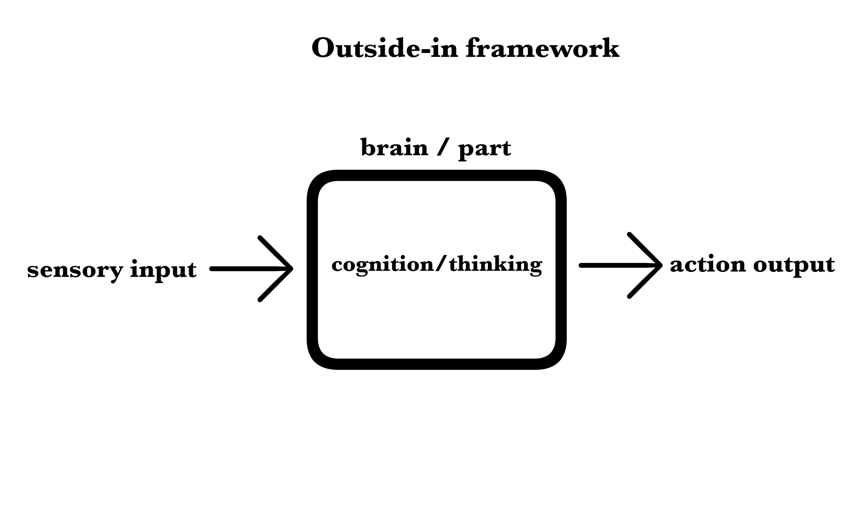
In this approach, the outside world conveys certain physical realities (sights, sounds, odors etc) which are accurately picked up by our senses and faithfully coded or represented in neuronal spikes which are then sent to the ‘cognitive’ / thinking part of the brain, namely the cortex.
The cortex or multiple cortical areas ‘process’ the sensory representations using algorithms that implement abilities like visual, auditory, olfactory attention, sense specific memories and other cognitive processing to finally generate an appropriate response to the input. This response is then executed by muscles or glands secreting hormones or, even, structures giving rise to feelings and / or emotions. This is the ‘outside-in’ framework in a nutshell.
original artwork
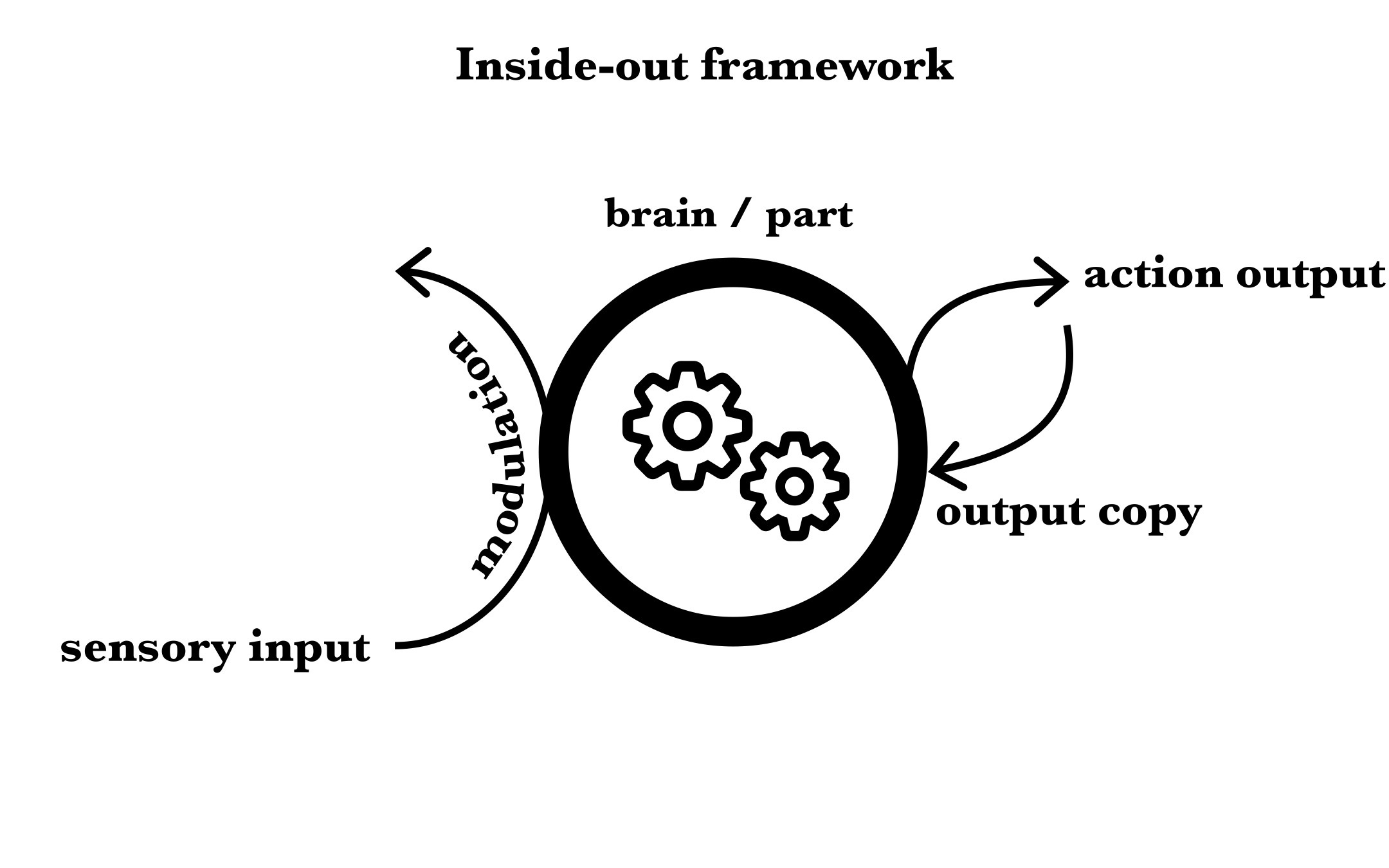
With over 50 years of state-of-the-art neurophysiological research on the hippocampus combined with an appreciation for the evolutionary and developmental history of the brain, Buzsáki strongly advocates for a very different kind of approach to studying the brain. He calls it the ‘inside-out view’ of the brain which I have attempted to sketch below and describe very briefly in general terms.
original artwork
The brain, or any of its sub-systems in general, are first and foremost a self-contained and self-interested network that is constantly active as indicated by the central hub with internal gearing. Sensory inputs modulate, as opposed to being encoded or represented faithfully by the sensory areas of the brain. This perturbs the ongoing activity of the cortical and sub-cortical networks, which then results in an action output (movement/secretion/feeling/emotion). Crucially, this action is copied back to the relevant cortical networks since they need to anticipate or predict the consequences of any action taken by the organism in response to their output.
How does one choose between these two very different frameworks for studying the brain? I don’t believe it is an open and shut case despite the way I have somewhat critically described the ‘outside-in’ approach above.
The differences and relative merits of both approaches are best appreciated by digging deep into the, at least, 70 year old quest to understand the hippocampus; its role in navigating physical space, and surprisingly, an apparently completely different capability: forming long-term memories. Nothing in philosophy or traditional psychology allows us to lump these two capabilities together and yet, clinical and experimental evidence suggests the hippocampus is deeply implicated in both these capabilities.
We have explored long-term memories a bit in earlier posts (see here and here). We convinced ourselves somewhat that our autobiographical self, our identity, is dependent on our long-term memories of events / episodes that happen to us. These memories are formally known as episodic memories8 in psychology and their formation and recall requires an intact hippocampus.
An easy way to think of episodic memories is what, where and when memories that happened to you.
One of my earliest memories (from decades ago) is of me falling down (what) with a full-sized, old, heavy, steel bicycle that I was pushing up our driveway (where), when I was perhaps 5 years old (when). My mother then rescued and comforted me, took me inside and gave me a warm glass of milk with a slice of bread. I can vividly recall feeling totally helpless as the bicycle fell and I can recall the smell and taste of that bread dipped in milk and how that felt so comforting. Indeed, I believe my lifelong habit of dipping bread in milky tea may have at least partially originated that day.
This is very remarkable, if you pause to think about this. We can remember things from the distant past that happened only once in our life. People remember select childhood episodes vividly even at the end of their lives. They can, decades later, recall the colors of clothing, the perfume someone was wearing, even how they themselves felt, at the exact moment when the incident happened. These significant moments leave lasting imprints on our life stories. There is good evidence to suggest our hippocampi are absolutely required for this instant formation of lifelong memories. So in that sense, the hippocampus has a role to play in constructing our autobiographical self.
Where and how is all this information stored in our brain? How can we recall so many aspects of an incident that happened years ago and never again? We only have rudimentary insight into these questions at this time.
We will dig into the over 70 year old scientific literature on the hippocampus and the even more exciting and conceptually sophisticated work being done these days over perhaps at least a couple of more posts.
Please do share your thoughts, objections, queries about this post. Also please circulate this post to any students who are interested in looking into cognitive neuroscience or AI work as a career. This field attracts as many people with engineering, computer science, mathematics physics backgrounds as people who have studied psychology, medicine or biology these days. It feels like a new dawn in neuroscience.


György Buzsáki, 2019, The Brain from Inside Out
William James 1890, The Principles of Psychology
Rodolfo Llinas, 2002, i of the vortex From Neurons to Self
Molnár, Z., Luhmann, H. J., & Kanold, P. O. (2020). Transient cortical circuits match spontaneous and sensory-driven activity during development. Science, 370(6514), eabb2153
Endel Tulving, 1985, Elements of episodic memory